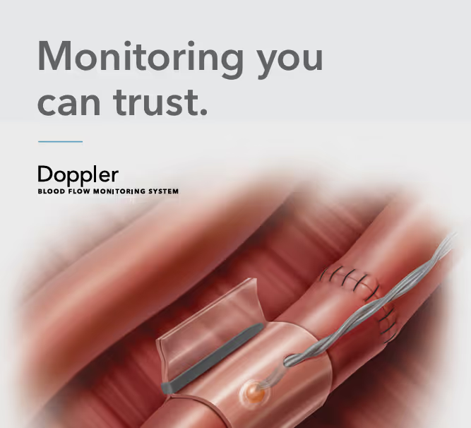
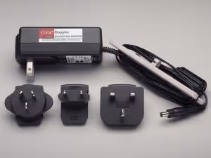
Doppler – Blood Flow Monitor


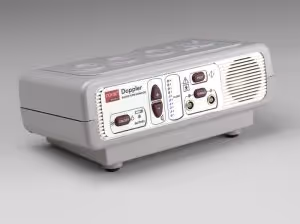

The Cook Doppler – Monitoring you can trust.
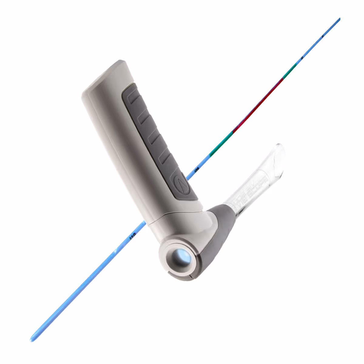
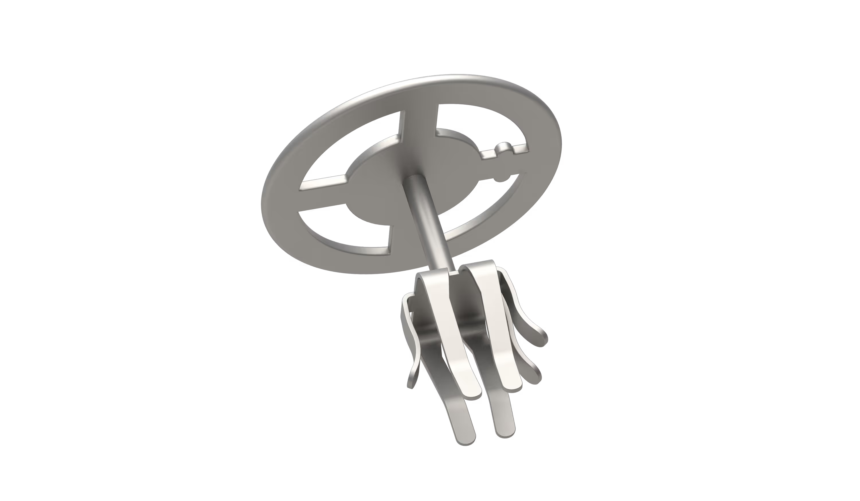
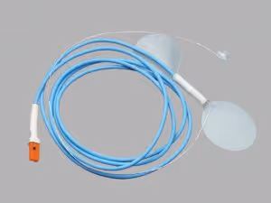
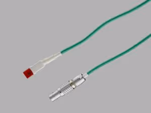
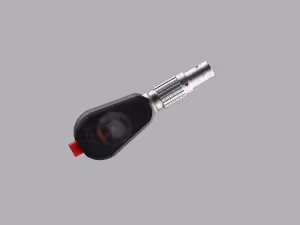
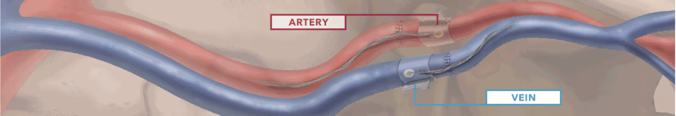
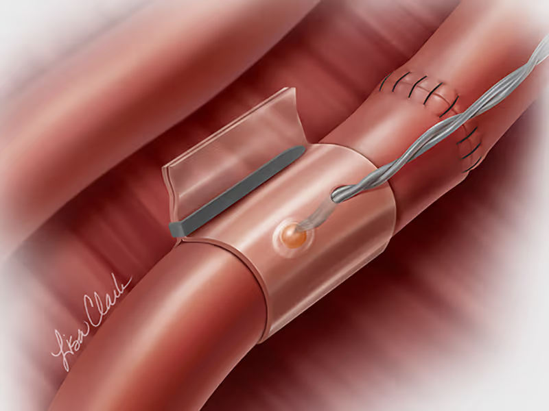
The Doppler monitor provides the surgeon with both audible (primary) and visual (secondary) feedback of blood flow when connected to the implantable Cook-Swartz Doppler Probes and extension cables. The system is very flexible, allowing for the effective monitoring via arteries and/or veins, end to end or end to side, proximal or distal to the anastomosis.
Another benefit of the device is its mobility, the device can be used in theatres and then moved with the patient into recovery and the ward. This enables constant monitoring and by allowing the team to see and hear the presence or absence of blood flow, the Doppler system can alert you to flap failure in time to perform a salvage procedure.
Clinical Evidence
Over twenty years of clinical evidence has established the Doppler Blood Flow Monitoring System can contribute to more favourable outcomes compared to conventional monitoring. The literature shows that the use of the Cook Doppler can:
Decrease returns to surgery – Because the Doppler system offers objective monitoring at an earlier stage, it has been associated with a decrease in returns to surgery 1.
Offer higher salvage rates – When compared with clinical monitoring alone, Doppler offers higher salvage rates 2,3 a systematic review and meta-analysis showed an increase of between 57% and 73% with the Doppler 2.
Offer lower failure rates – It has also been shown that using an implantable Doppler may reduce the number of flap failures by at least 37% 2.
Demonstration
To arrange a meeting or demonstration please contact us via the Info Request form below.
Video
A set of video’s covering many aspects the Cook Doppler can be found via the Video Library tab at the top of this page.
These videos include;
- Doppler Blood Flow Monitor IFU
- Surgical placement of the probe
- Intraoperative troubleshooting
- Postoperative troubleshooting
- Probe removal
You can also learn more about the system via our online brochure, that focuses on the use of the system within head and neck. Please see the PDF tab at the top of this page.
References:
1. Wax MK. The role of the implantable Doppler probe in free flap surgery. Laryngoscope. 2014;124(Suppl 1):S1-S12.
2. Chang TY, Lee YC, Lin YC, et al. Implantable Doppler probes for postoperatively monitoring free flaps: efficacy. A systematic review and meta-analysis. Plast Reconst Surg Glob Open. 2016;4(11):e1099.
3. Lenz Y, Gross R, Penna V, et al. Evaluation of the implantable Doppler probe for free flap monitoring in lower limb reconstruction. J Reconstr Microsurg. 2018;34(3):218-226.
Tel: 01264 332172
Email: info@CCMed.co.uk
We hope you found the videos of interest. To learn more or request a demonstration please contact us on 01264 332172, email info@ccmed.co.uk or use our contact form.