Biodesign Advanced Tissue Repair – handle with ease, repair with confidence
- Providing a strong seal
- Excellent handling
- Sutured/Sutureless fixation


Biodesign® is a platform technology behind numerous tissue-repair products that span multiple medical specialties. Biodesign is natural extracellular matrix (ECM) derived from porcine small intestinal submucosa (SIS). The ECM is a complex latticework of proteins and structural molecules that helps guide the growth of cells.
Biodesign’s proprietary processing methodology decellularizes the SIS material while preserving natural matrix molecules such as collagen, proteoglycans, and glycosaminoglycans. The result is a scaffold that, when implanted, provides a location for host cells to infiltrate and remodel into vascularized tissue.
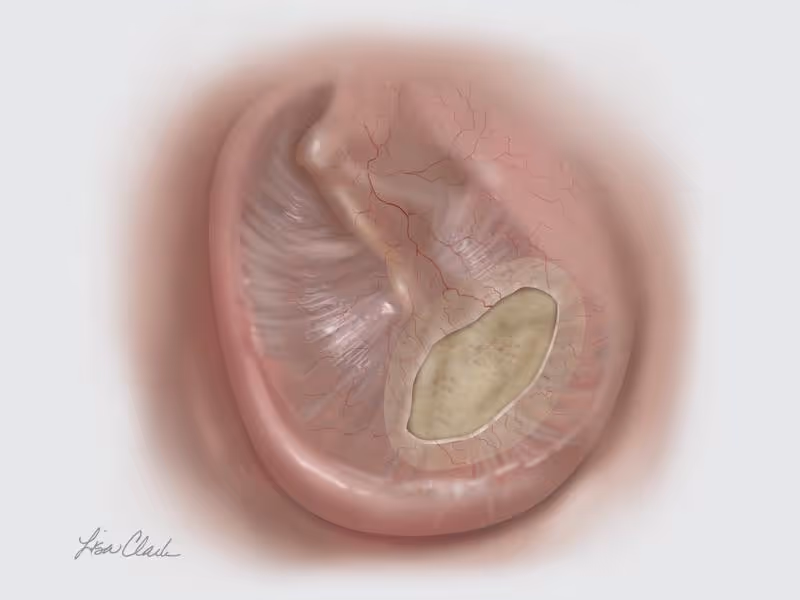
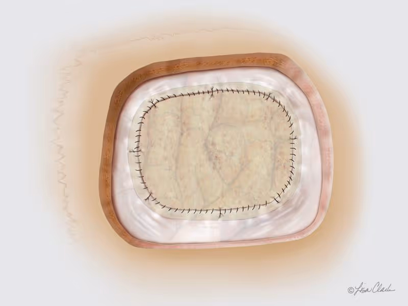
Biodesign Duraplasty Graft is used to repair the dura mater following surgical procedures (e.g. tumour resection). In other cases, the graft may be used to treat existing CSF leaks such as those resulting from trauma or sinus surgery.1,4
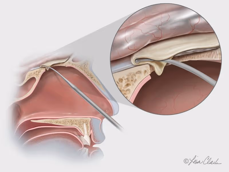
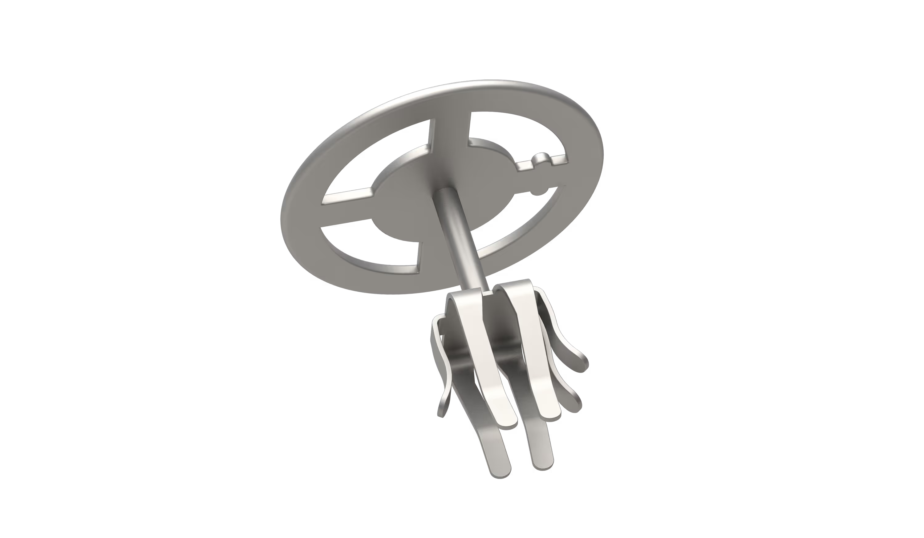
Biodesign material is easy to manipulate, doesn’t swell with hydration, and doesn’t adhere to itself when folded. It can be secured in place with or without sutures, depending on clinician preference.4
It shares the same characteristics as the rest of the Biodesign range and in this indication it is used as an onlay graft that can be secured in place with or without sutures, depending on clinician preference.
Tel: 01264 332172
Email: info@CCMed.co.uk
We hope you found the videos of interest. To learn more or request a demonstration please contact us on 01264 332172, email info@ccmed.co.uk or use our contact form.